M. hyo
Mycoplasma hyopneumoniae infections
Mycoplasma hyopneumoniae (M. hyo) is the primary pathogen causing enzootic pneumonia (EP), a chronic respiratory disease in pigs, and one of the primary agents involved in the porcine respiratory disease complex (PRDC) (Maes et al. 2017).
EP is characterized by a chronic and persistent, dry, hacking cough, decreased growth rate and feed conversion ratio (Sibila et al. 2009), typically with no or low mortality unless accompanied by a secondary infection.
PRDC develops because of co–infections of both bacterial and viral pathogens, especially porcine reproductive and respiratory syndrome virus (PRRSV), swine influenza virus (SIV), and porcine circovirus type 2 (PCV2) (Sibila et al.2009).
PRDC can result in increased mortality and severe performance losses. The major threat for the farm economy is represented by the decrease in average daily gain (ADG) the daily weight gain and eventual increased medication cost. Infection with M. hyo often appears to have a subclinical course, where only the growth performance is reduced.
M.hyo bacteria
M.hyo is one of the smallest self-replicating microorganisms with a missing cell wall. The pathogen is unable to survive long outside its host in the environment, but in aerosols its survival time increases as it can remain infectious for up to 31 days in water at 2-7°C (Villareal 2010). M.hyo can travel in the air up to 4-5 km via aerosols.
The clinical outcome of M.hyo infections is determined by different factors, such as housing conditions, management practices, co‐infections and by virulence differences among M.hyo isolates. Significant variation has been observed among different Mycoplasma hyopneumoniae isolates at the antigenic, proteomic, transcriptomic, pathogenic, and genomic levels, as well as in their virulence (Meyns 2007; Vicca 2003; Betlach 2019). Although the mechanisms of pathogenesis are not yet fully understood, research has identified several contributing factors. These include specific proteins—particularly adhesins—as well as genes involved in metabolic processes and bacterial growth, all of which appear to play a role in determining the organism’s virulence.
High virulent strains induce faster and more severe symptoms than low virulent strains (Villareal 2009). Low virulent strains also induce later seroconversion. However, previous exposure to the low virulent strain doesn’t reduce the lesions due to the subsequent high virulent M.hyo infection.
Co‐infection with more than one strain in a pig or batch of pigs might result in more severe lung lesions (Michiels et al., 2017).
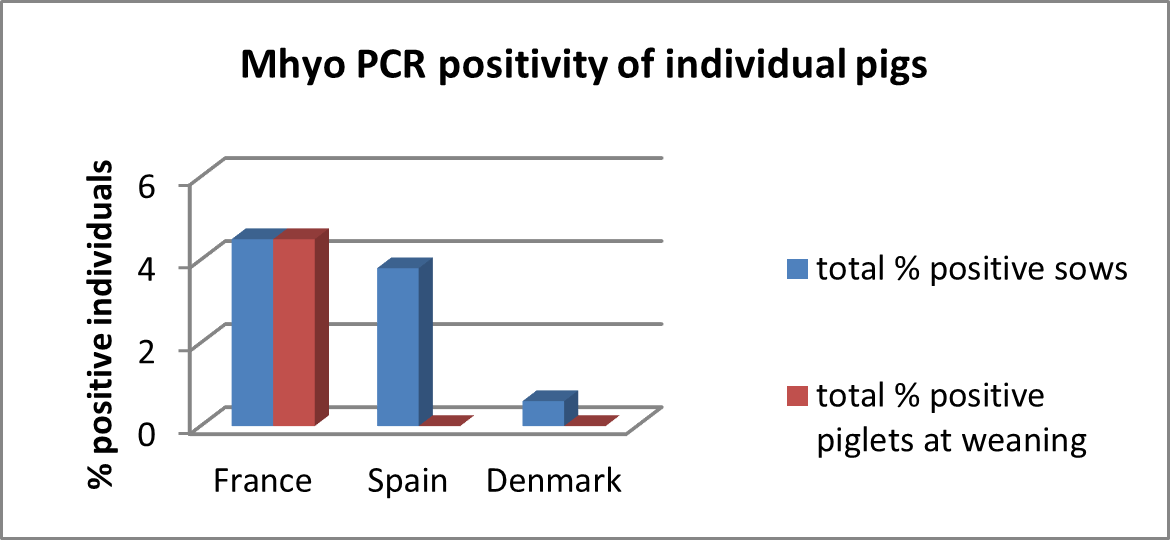
Prevalence of nPCR positive results, average number of different strains, severity of Mycoplasma-like lesions ±SD, prevalence pneumonia, fissures and pleurisy expressed in percentages. (Michiels et al., 2017)
1 = batches with only one strain detected, 2 = batches with 2–6 different strain and 3 = batches with ≥7 strains detected

Disease
Mycoplasma hyopneumoniae (M. hyo) is the primary pathogen causing enzootic pneumonia (EP), a chronic respiratory disease in pigs, and one of the primary agents involved in the porcine respiratory disease complex (PRDC) (Maes et al. 2017). Pneumonia induced by M. hyo is considered one of the most widespread and important chronic diseases in pigs. The prevalence is estimated to be as high as 80% in the global pig population (Batista 2006). In countries with advanced pig-farming practices, between 30% and 80% of slaughtered pigs exhibit lung lesions characteristic of Mycoplasma hyopneumoniae infection (Scott A. Dee, DVM, MS, PhD, DACVM, Nearly all (more than 99%) of commercial swine herds have mycoplasmal pneumonia in United States. In the Canadian swine industry, Mycoplasma hyopneumoniae (M. hyo) remains one of the most prevalent and economically significant respiratory pathogens.
Reservoir of the infection:
Sows and piglets in breeding herds are considered the reservoir of M.hyo infections for the entire production system. Circulation of M.hyo is thought to occur among existing sows and be transmitted to incoming gilts, which are capable of maintaining the pathogen within the farm and are responsible for the majority of bacterial shedding to newborn pigs. Infection with M. hyo has a long duration, reaching up to 240 days (Pieters 2009), complicating the already slow disease transmission scenario observed in sow herds. Transmission occurs through nasal secretions (nose-to–nose) and coughing secretions.
Prevalence:
Positive rates vary across age groups, with the highest levels of circulation typically observed in growing-finishing pigs. In contrast, the prevalence among weaned pigs remains very low on most conventional farms (Krejci, 2019).
Typical clinical presentation:
Typical clinical picture of enzootic pneumonia includes persistent, dry cough, anorexia, and dyspnea. Dynamics and duration of coughing are important for the estimation of the impact of EP on growth retardation. It is difficult to assess the economic effect of mycoplasmal pneumonia because of the multifactorial origin of PRDC. Enzootic pneumonia has been associated with a 17% reduction in average daily weight gain and a 14% decline in feed efficiency in affected herds (Straw, 1989).
Impact on performance:
Research shows that when healthy pigs free from M. hyo were mixed into M. hyo positive herds and were thus exposed to the natural infection, their performance was lower. The ADG of pigs with a non-complicated mycoplasmal pneumonia decreases by more than 6-g/day compared to those that remain free from M.hyo , after adjusting for herd, pen, weight and sex (Rautiainen et al. 2000).
Diagnostic:
The presence of the infection is usually confirmed by M.hyo specific seroconversion or by detecting the pathogen by PCR in the laryngeal swabs (Pieters et al. 2017, Sibila et al. 2009). M. Hyo Infected lung tissue develops consolidation and catarrhal broncho-pneumonia characterized by purple to grey regions of meaty aspect. The consolidation can be observed from 3-12 weeks post infection. The lesions are mainly localized in the apical and cardiac lobes, as well as in the anterior part of the diaphragmatic lobes and in the intermediate lobe. Lesions resolve after 12 to 14 weeks with formation of interlobular fissures (Maes et al. 2008). Considering the chronic nature of these lesions, bronchopneumonia with the cranioventral consolidation of lungs is very indicative of EP in slaughter pigs.
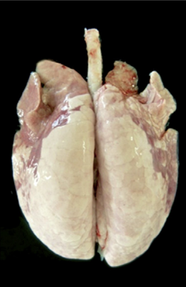
Example of a lung with lesions derived from an experimental M.hyo inoculation (Garcıa-Morante, 2016).