PCV2
Porcine Circovirus (PCV2) infections
Porcine circovirus type 2 (PCV2) is ubiquitous in Canada and present in virtually all swine herds worldwide. The discovery of the virus was associated with the first cases of post‑weaning multisystemic wasting syndrome (PMWS) in piglets. The disease was first observed in Canada in 1991 as an unclassified wasting syndrome, accompanied by high mortality after weaning.
This new syndrome was characterized by:
- weight loss
- respiratory distress,
- pale skin,
as well as typical pathological lesions such as:
- lymphadenopathy,
- interstitial pneumonia,
- and hepatitis.
The name PMWS, along with a detailed description of the syndrome, was published in 1996. In 1998, a genetically and antigenically distinct porcine circovirus was identified as the cause of the disease and was designated porcine circovirus type 2 (PCV2).
PCV2 causes immunosuppression, making pigs more susceptible to secondary diseases.
Difference between PCV2 and PCV1:
Unlike PCV2, porcine circovirus type 1 (PCV1), isolated earlier, is considered non-pathogenic and has been found in pigs showing no clinical signs.
However, serological studies have demonstrated that PCV2 had already been circulating asymptomatically in swine populations for at least 25 years before the first reported case of PCV2-associated PMWS.
The spread of new, more virulent PCV2 strains across global swine operations is believed to be linked to the movement of breeding animals, semen, or other management practices (Krakowka 2012).
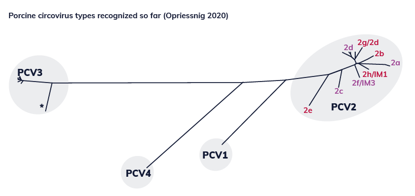
Emergence of new circovirus types:
Since then, other types have been identified, including:
- PCV3 (Palinsky 2017)
- PCV4 (Zhang 2019)
These new types were distinguished based on differences in the ORF2 gene (open reading frame 2).
Porcine circovirus type 2 (PCV2) has become one of the most important swine viruses and is associated with several pathological syndromes in nursery piglets, and even more so in growing‑finishing pigs.
- In 2002, the term “porcine circovirus diseases” (PCVD) was proposed to include both clinical and subclinical forms.
- In 2006, the term PCVAD (porcine circovirus associated diseases) was used for the first time, mainly in North America.
- In 2012, Segalés proposed a distinction between:
- PCV2 subclinical infections,
- and the different clinical forms associated with PCV2.
In pigs, from weaning to slaughter, the following syndromes are described.
Porcine circovirus diseases (PCVD)
PCV2 subclinical infection (PCV2-SI)
- Despite the absence of clinical signs, decreased average daily gain and higher percentage of runts (lower birth weight) can be observed and consequently improved by vaccination.
- No or minimal histopathological lesions in tissues (mainly lymphoid).
- None or low IHC (Immuno Histo Chemistry). Low amount of PCV2 in few (lymphoid) tissues (qPCR <105-106 / g tissue).
PCV2 systemic disease (PCV2-SD)
- Wasting, weight loss/emaciation, enlarged lymph nodes, and paleness of skin/jaundice. Respiratory signs can include coughing and tachypnea, and diarrhea can be observed as a digestive symptom. Note that clinical signs can vary significantly for this disease. Morbidity is usually 4-30%, mortality rates can reach 20%.
- Moderate to severe lymphocyte depletion with granulomatous inflammation of lymphoid tissues (plus granulomatous inflammation in a number of other tissues).
- IHC – moderate to high in lymphoid and other tissue; (qPCR >106 / g tissue).
Porcine dermatitis and nephropathy syndrome (PDNS)
- Dark red papules and macules on skin, mainly in hind limbs and perineal areas. Hemorrhagic and necrotizing skin lesions and/or swollen and pale kidneys with generalized cortical petechia.
- Systemic necrotizing vasculitis and necrotizing and fibrinous glomerulonephritis.
- None or low IHC in lymphoid tissue, (qPCR<106/g tissue).
PCV2 enteric disease (PCV2-ED)
- Diarrhea.
- Granulomatous enteritis and lymphocyte depletion with granulomatous inflammation in Peyer’s patches but no other lymphoid tissues.
- Moderate to high IHC in intestinal mucosa/Peyer’s patches.
PCV2 lung disease (PCV2-LD)
- Respiratory distress, dyspnea.
- Interstitial or broncho-interstitial pneumonia, mild to- severe necrotizing and ulcerative bronchiolitis or proliferative necrotizing pneumonia in the absence of lymphoid lesions as indicated for PCV2-SD.
- Moderate to high IHC in the lung.